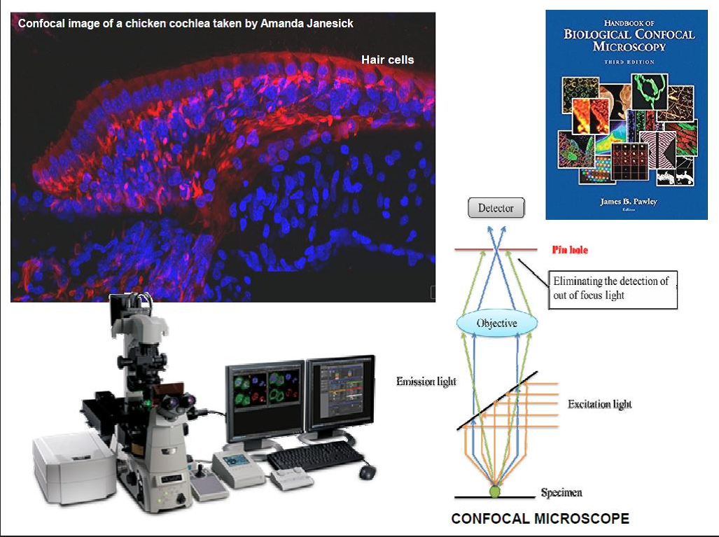
Fluorescent G proteincoupled receptors (GPCRs) G proteincoupled receptors (GPCRs) are the largest class of transmembrane proteins and the targets for almost half of the clinical drugs in the market today. UC San Diego Bioengineering: Applying engineering principles to scientific discovery and technology innovation to improve health, quality of life and to train future biotechnology leaders. 8094 MRC Laboratory of Molecular Biology, Neurobiology Postdoctoral Scientists Postdoctoral ScientistStarting Salary 30, 464 to 33, 305 per annumMRC Laboratory of Molecular Biology, Cambridge, UKThe MRC Laboratory of Molecular Biology is a world leader in biological research, with an expanding scientific programme dedicated to understanding gene function in health and disease. Genetic Laboratory Testing: Genetic Disease Gene Tests. MEDICAL GENETICS TESTS Medical Genetics Laboratories, Department of Molecular and Human Genetics, Baylor College of Medicine Multimedia Medical Genetics Tests Database (Text Images). For more information see Medical Genetics Laboratories or the Department of Molecular and Human Genetics University of Alberta. How poker and other games help artificial intelligence evolve. Michael Bowling's research in artificial intelligenceand how it intersects with games and machine learninghas put him at the forefront of the rapidly evolving field. Buy Handbook of Biological Confocal Microscopy on Amazon. com FREE SHIPPING on qualified orders The field of fluorescence microscopy is experiencing a renaissance with the introduction of new techniques such as confocal, multiphoton, deconvolution, and total internal reflection, especially when coupled to advances in chromophore and fluorophore technology. INTRODUCTION OVERVIEW Biology as a science deals with the origin, history, process, and physical characteristics, of plants and animals: it includes botany, and zoology. A study of biology includes the study of the chemical basis of living organisms, DNA. Other related sciences include microbiology and organic chemistry. Do topo para baixo, um MET consiste de uma fonte de emisso, a qual pode ser um filamento de tungstnio, ou uma fonte de hexaboreto de lantnio (LaB 6). [30 Para tungstnio, esta ser da forma de um filamento de um grampo de cabelo, ou um filamento em forma de pequena espiga. Microscopy is the technical field of using microscopes to view objects and areas of objects that cannot be seen with the naked eye (objects that are not within the resolution range of the normal eye). There are three wellknown branches of microscopy: optical, electron, and scanning probe microscopy. Optical microscopy and electron microscopy involve the diffraction, reflection, or refraction. This web page explains how a confocal microscope works; I've tried to make this explanation not too technical, although for certain parts I've included some details for people who know more optics. A Manual of Applied Techniques for Biological Electron Microscopy. Dykstra, 1993, 2nd Printing, 272pp, spiral bound, ISBN. Research news, the latest microscopes and accessories, meetings, short courses, and webinars for microscopists. Welcome to the third edition of the Handbook of Biological Statistics! This online textbook evolved from a set of notes for my Biological Data Analysis class at the University of Delaware. My main goal in that class is to teach biology students how to choose the appropriate statistical test for a particular experiment, then apply that test and interpret the results. Images in fluorescence microscopy are inherently blurred due to the limit of diffraction of light. The purpose of deconvolution microscopy is to compensate numerically for this degradation. Ultrafiltration (UF) is one of the best options for both onestage and as part of multistage water and wastewater purification. This review summarises the known facts about the fouling processes and cleaning procedures and details of the most successful physical and chemical cleaning combinations. Confocal microscopy, most frequently confocal laser scanning microscopy (CLSM) or laser confocal scanning microscopy (LCSM), is an optical imaging technique for increasing optical resolution and contrast of a micrograph by means of using a spatial pinhole to block outoffocus light in image formation. Capturing multiple twodimensional images at different depths in a sample enables the. The Handbook of Biomedical Nonlinear Optical Microscopy provides comprehensive treatment of the theories, techniques, and biomedical applications of nonlinear optics and microscopy for cell biologists, life scientists, biomedical engineers, and clinicians. The chapters are separated into basic and advanced sections, and provide both textual and graphical illustrations of all key concepts. The Microscopy ListServer Sponsor: The Microscopy Society of America 33rd Scottish Microscopy Symposium Wednesday 9th November 2005, Hunter Halls, University of Glasgow, Glasgow Un microscope confocal, appel plus rarement microscope monofocal, est un microscope optique qui a la proprit de raliser des images de trs faible profondeur de champ (environ 400 nm) appeles sections optiques. En positionnant le plan focal de lobjectif diffrents niveaux de profondeur dans lchantillon, il est possible de raliser des sries dimages partir.


